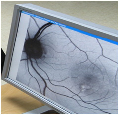
Angiography: Fluorescein or (Indocyanine green) ICG is injected into the brachial vein and fundus camera is used for imaging the back of the eye. This test is used to check for retinal and choroidal blood flow. Fluorescein is commonly used to examine the retinal vessels and ICG to examine the choroidal and deeper vessels. Fluorescein angiography is more commonly used for the evaluation of diabetic retinopathy, obstructive vascular disease such as arterial obstruction or retinal vein evaluation. ICG is used to screen for blood in the macula, such as age-related degenerative warts, both of which have very few side effects and can be used safely. Allergic reactions may rarely occur in some people. ICG is prohibited in people with iodine sensitization. Some people develop jaundice on the skin or eyes for up to 24 hours after injection, causing urine to turn orange.
OCT or Optic Coherence Tomography: A new technique that provides ophthalmologists with valuable information from retinal layers using high-resolution tomographic sections. Therefore, it is used to diagnose and track many retinal diseases such as macular holes, macular edema, macular degeneration, diabetic retinopathy and glaucoma. Since this technique uses a light source, there is no need for eye contact and it takes only a few seconds.